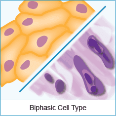
One interesting study is called, “Proteoglycans in human malignant
mesothelioma. Stimulation of their
synthesis induced by epidermal,
insulin and platelet-derived growth factors involves receptors with
tyrosine kinase activity” – Biochimie Volume 81, Issue 7, July 1999,
Pages 733-744 – by Alexandra Syrokoua, George N. Tzanakakisb, Anders
Hjerpeb and Nikos K. Karamanosa - Department of Chemistry, University of
Patras, 261 10 Patras, Greece.
Here is an excerpt: “Abstract -
Identification of proteoglycans in two human malignant mesothelioma cell
lines, one with epithelial differentiation and the other with
fibroblast-like phenotype, and the effects of epidermal (EGF),
insulin-like (IGF-I) and platelet-derived (PDGF-BB) growth factors on
the synthesis of hyaluronan (HA) and proteoglycans (PGs) were studied.
Both cell lines synthesize HA and PGs: these last were recovered both as
secreted and cell-associated compounds.
Chondroitin sulfate (CS)
containing PGs are mainly organized as versican in the extracellular
medium and as thrombomodulin and syndecan in the cell membrane. Heparan
sulfate (HS) containing PGs are mainly in the form of perlecan in the
culture medium, whereas cell-associated HSPGs were recovered mainly as
syndecan-1, -2 and -4. Receptors for EGF, IGF-I and PDGF-BB were
identified in both cell lines. In addition to cell proliferation, these
growth factors stimulated the synthesis of HA and PGs, the pattern of
stimulation being unique for each of them and depending on the cell
phenotype.
EGF increased the synthesis of HA and PGs. IGF-I showed
similar stimulatory effects on the synthesis of CSPGs, whereas higher
amounts were needed to influence the synthesis of HA and HSPGs, the
latter only being stimulated in the epithelial cell line. PDGF-BB
stimulated the synthesis of HA, HSPGs and CSPGs at low concentrations,
while the stimulatory effect was abolished at higher levels. Incubation
with genistein inhibited the HA and PG synthesis induced by growth
factors in a mode depending on both growth factor and genistein
concentrations. The results clearly suggest that the stimulatory effects
of EGF, IGF-I and PDGF-BB on matrix synthesis, expressed as
proteoglycan synthesis, are mediated via receptor-growth factor
complexes and the protein tyrosine kinase intracellular pathway.
Another study is called, “Value of E-cadherin and N-cadherin immunostaining in the diagnosis of Mesothelioma” by ORDONEZ Nelson G. 2, Allée du Parc de Brabois F-54514 Vandoeuvre-lès-Nancy – Cedex France. Here is an excerpt: “Abstract - Distinguishing between epithelioid mesothelioma and pulmonary adenocarcinoma involving the pleura can be difficult on routine histological preparations. This differential diagnosis can be greatly facilitated by using immunohistochemical markers. E-cadherin and N-cadherin are among the newly described markers that have been proposed as potentially useful in the diagnosis of mesothelioma. E-cadherin and N-cadherin are members of the cadherin family of calcium-dependent cell adhesion molecules that play an important role in the embryogenic development and maintenance of normal tissue.
Another study is called, “Value of E-cadherin and N-cadherin immunostaining in the diagnosis of Mesothelioma” by ORDONEZ Nelson G. 2, Allée du Parc de Brabois F-54514 Vandoeuvre-lès-Nancy – Cedex France. Here is an excerpt: “Abstract - Distinguishing between epithelioid mesothelioma and pulmonary adenocarcinoma involving the pleura can be difficult on routine histological preparations. This differential diagnosis can be greatly facilitated by using immunohistochemical markers. E-cadherin and N-cadherin are among the newly described markers that have been proposed as potentially useful in the diagnosis of mesothelioma. E-cadherin and N-cadherin are members of the cadherin family of calcium-dependent cell adhesion molecules that play an important role in the embryogenic development and maintenance of normal tissue.
Although several investigations have indicated that
immunostaining for these markers can be useful in discriminating between
mesotheliomas and adenocarcinomas, others have not confirmed this
observation. In an attempt to resolve this controversy, the present
study investigated 31 epithelioid mesotheliomas and 29 pulmonary
adenocarcinomas for E-cadherin and N-cadherin expression using the 5H9,
HECD-1, and clone 36 anti-Ewadherin antibodies, and the 3B9 and clone 32
anti-N-cadherin antibodies. Among the mesotheliomas, 68% reacted with
the clone 36, 52% reacted with the HECD-1, and 19% reacted with the 5H9
anti-Ecadherin antibodies, and 74% reacted with the 3B9 and 71% reacted
with the clone 32 anti-N-cadherin antibodies.
Of the adenocarcinomas,
93% stained with the done 36, 90% reacted with the HELD-1, and 90%
reacted with the 5H9 anti-Ecadherin antibodies, 45% reacted with the
clone 32 and 34% reacted with the 3B9 anti-N-cadherin antibodies. Based
on the frequent strong reactivity with adenocarcinomas but not with
mesotheliomas, it is concluded that only the 5H9 anti-Ecadherin antibody
may have some utility in discriminating between epithelioid pleural
mesotheliomas and pulmonary adenocarcinomas. The causes of the disparate
results reported in the literature on the value of E-cadherin and
N-cadherin immunostaining in distinguishing between mesotheliomas and
pulmonary adenocarcinomas are unclear, but a significant factor appears
to be differences in the reactivity of the antibodies used.”
Another study is called, “The value of Wilms tumor susceptibility gene 1 in cytologic preparations as a marker for malignant Mesothelioma” by Jonathan L. Hecht M.D., Ph.D., Benjamin H. Lee M.D., Ph.D., Jack L. Pinkus Ph.D., Geraldine S. Pinkus M.D., - Cancer Cytopathology Volume 96, Issue 2, pages 105–109, 25 April 2002. Here is an excerpt: “Abstract - It has been shown that detection of the Wilms tumor susceptibility gene 1 protein (WT1) has diagnostic utility in the distinction of mesothelioma from adenocarcinoma in tissue sections of pleural tumors. This immunohistochemical study evaluates the effectiveness of WT1 as a marker for malignant mesothelioma in paraffin sections of cell block preparations derived from effusion specimens. METHODS The authors evaluated 111 cell blocks for WT1 immunoreactivity, including 14 mesotheliomas and 97 metastatic adenocarcinomas from various sites.
Another study is called, “The value of Wilms tumor susceptibility gene 1 in cytologic preparations as a marker for malignant Mesothelioma” by Jonathan L. Hecht M.D., Ph.D., Benjamin H. Lee M.D., Ph.D., Jack L. Pinkus Ph.D., Geraldine S. Pinkus M.D., - Cancer Cytopathology Volume 96, Issue 2, pages 105–109, 25 April 2002. Here is an excerpt: “Abstract - It has been shown that detection of the Wilms tumor susceptibility gene 1 protein (WT1) has diagnostic utility in the distinction of mesothelioma from adenocarcinoma in tissue sections of pleural tumors. This immunohistochemical study evaluates the effectiveness of WT1 as a marker for malignant mesothelioma in paraffin sections of cell block preparations derived from effusion specimens. METHODS The authors evaluated 111 cell blocks for WT1 immunoreactivity, including 14 mesotheliomas and 97 metastatic adenocarcinomas from various sites.
RESULTS Nuclear reactivity for WT1 was observed in all samples of
mesothelioma. However, only 22 of 97 samples (23%) of metastatic
adenocarcinoma, nearly all of which were of ovarian origin (91%),
exhibited nuclear reactivity for WT1. In 14 other samples (most of
pulmonary derivation), WT1 staining restricted to the cytoplasm was
observed for some tumor cells and was regarded as nonspecific.
CONCLUSIONS - Based on this staining profile, WT1 represents an
effective marker for mesotheliomas in cell block preparations and can
aid in its distinction from pulmonary adenocarcinoma. In assessment of
effusion specimens with metastatic carcinoma, nuclear reactivity for WT1
is highly suggestive of an ovary primary tumor.
Wilms tumor susceptibility gene 1 is a tumor suppressor gene that initially was identified due to its deletion or mutation in Wilms tumors. Monoclonal antibodies to its protein product, WT1, were developed subsequently, and it was found that they had diagnostic utility not only in the identification of Wilms tumors and desmoplastic small round cell tumors1, 2 but also in the distinction of mesothelioma from adenocarcinoma in pleural tumors.3, 4 This immunohistochemical study evaluates the diagnostic utility of WT1 as a marker for malignant mesothelioma in paraffin sections of cell block preparations derived from effusion specimens. Cancer (Cancer Cytopathol) 2002;96:000–000.
Wilms tumor susceptibility gene 1 is a tumor suppressor gene that initially was identified due to its deletion or mutation in Wilms tumors. Monoclonal antibodies to its protein product, WT1, were developed subsequently, and it was found that they had diagnostic utility not only in the identification of Wilms tumors and desmoplastic small round cell tumors1, 2 but also in the distinction of mesothelioma from adenocarcinoma in pleural tumors.3, 4 This immunohistochemical study evaluates the diagnostic utility of WT1 as a marker for malignant mesothelioma in paraffin sections of cell block preparations derived from effusion specimens. Cancer (Cancer Cytopathol) 2002;96:000–000.


Post a Comment